
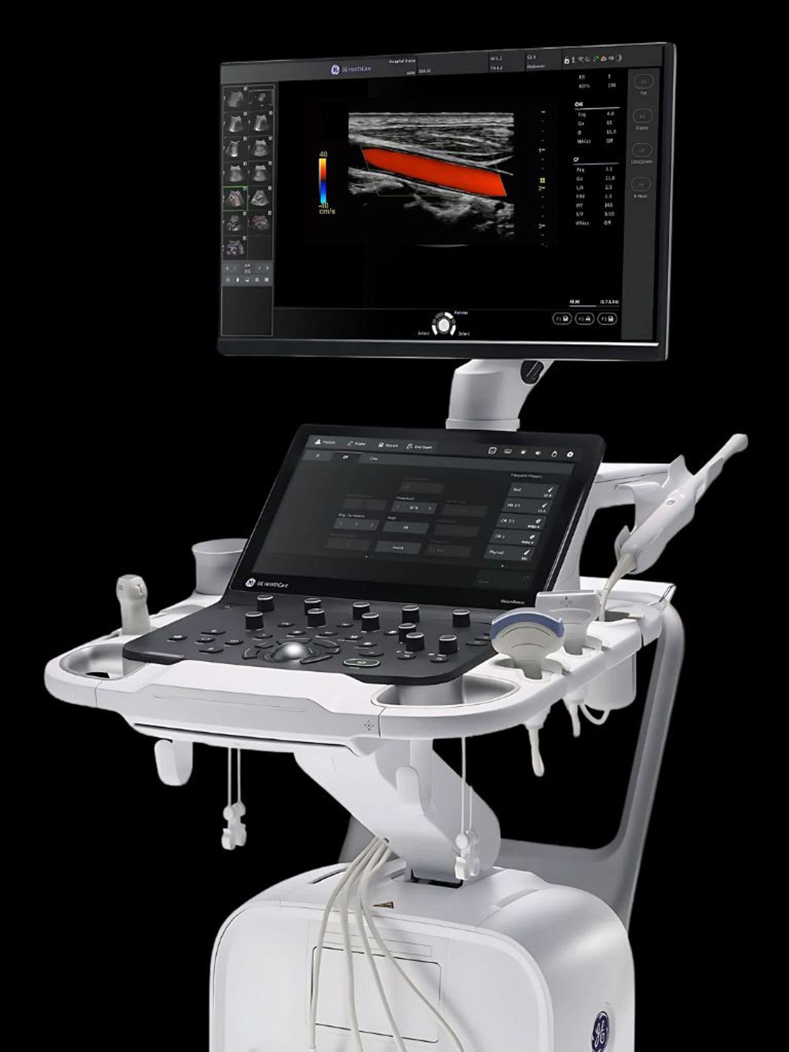
Certified Preowned GE Ultrasound Systems
Same GE equipment. Same GE warranty. 30-50% less than new. Every system is inspected, tested, and certified before it ships.
The same GE imaging. Lower capital investment.
A certified preowned GE system delivers the exact same image quality as a factory-new unit. The difference is your practice keeps $30,000-$60,000 in working capital.
Save 30-50% vs. new
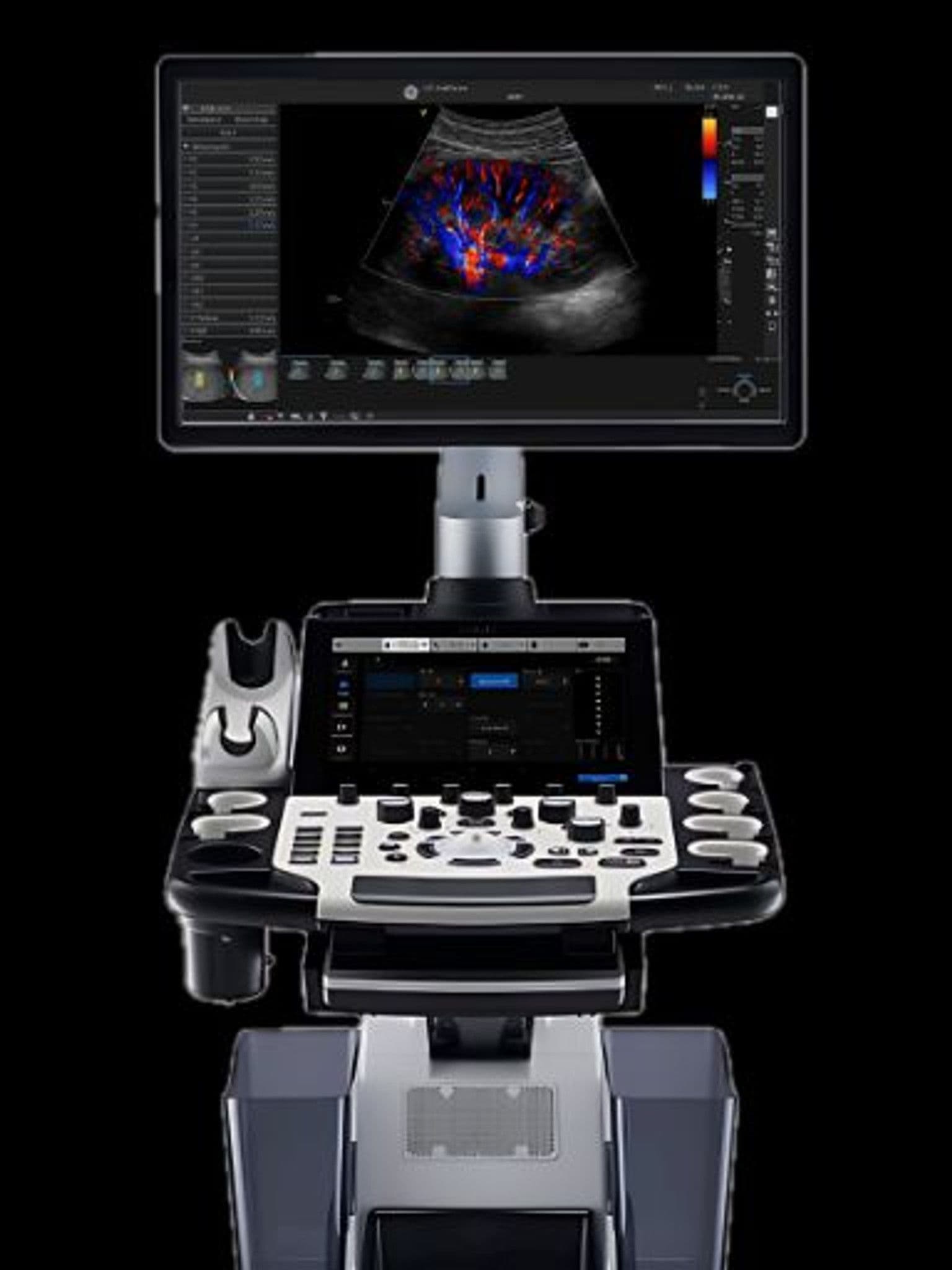
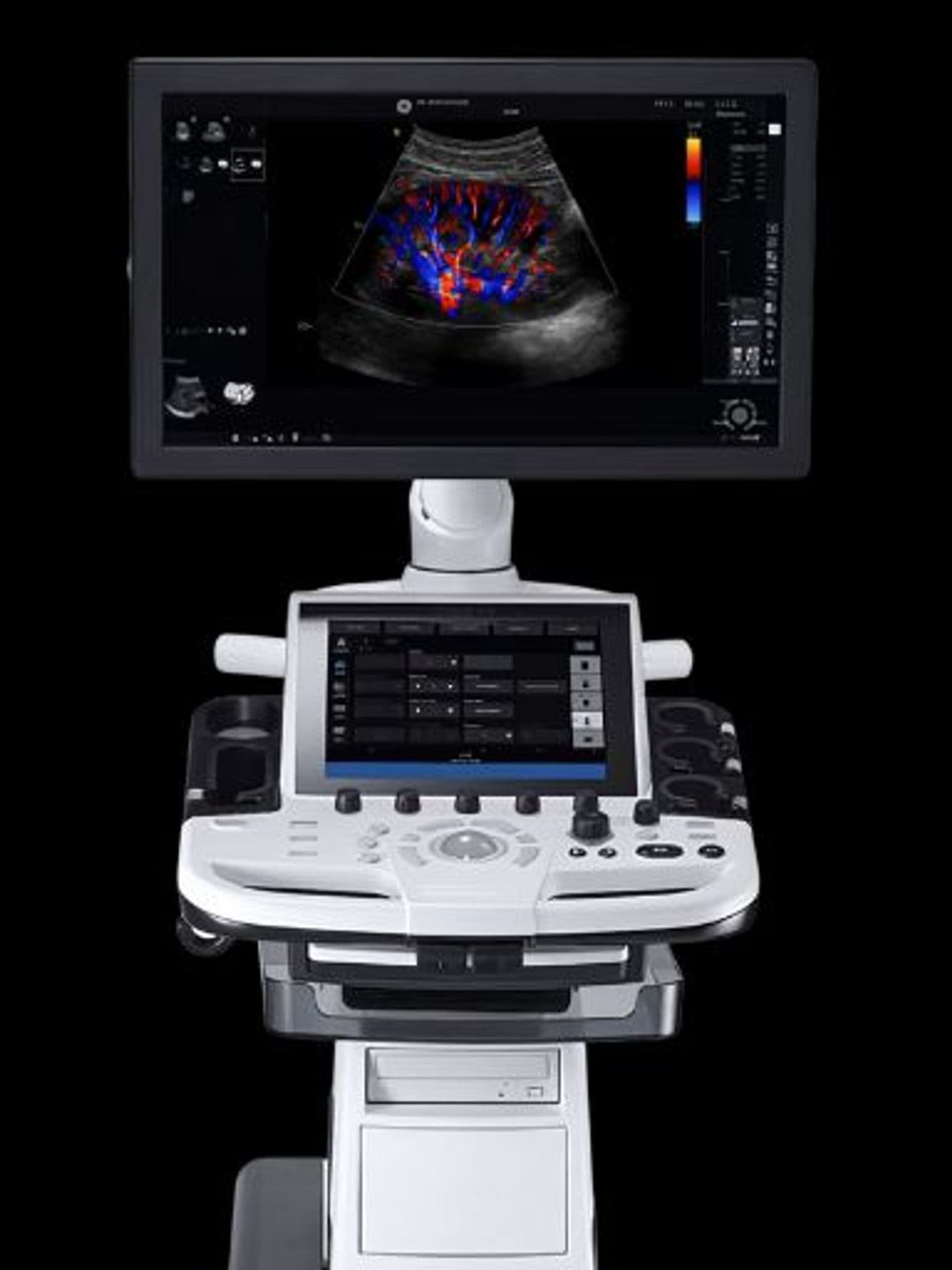
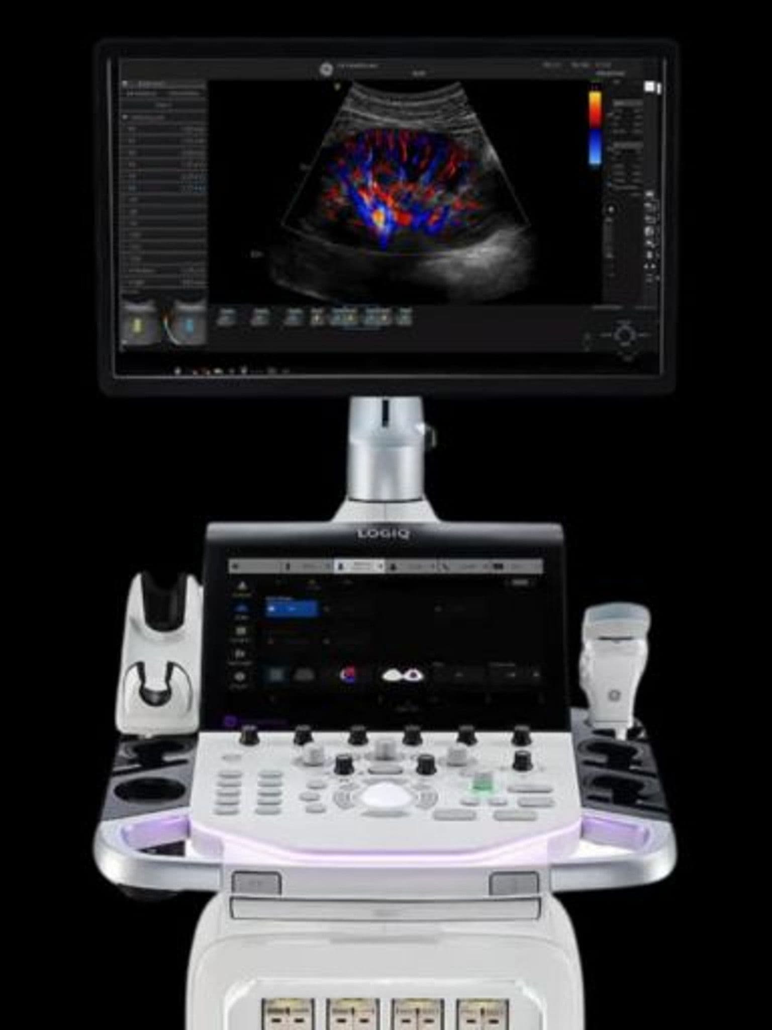
A preowned Vivid or LOGIQ system costs significantly less than new — without sacrificing the image quality or clinical capability your patients depend on.
Inspected and certified
Every preowned system is inspected by qualified technicians, tested against GE's original performance specifications, and updated with current software before delivery.
Full GE warranty included
Preowned systems purchased through Standard Ultrasound ship with GE warranty coverage. Same service network, same technicians, same response times as new.
30-day trial period
Use the system in your practice for 30 days. Scan real patients, test every mode. If it doesn't meet your clinical needs, we pick it up at no cost.
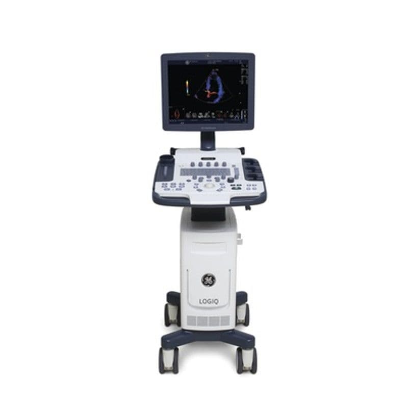
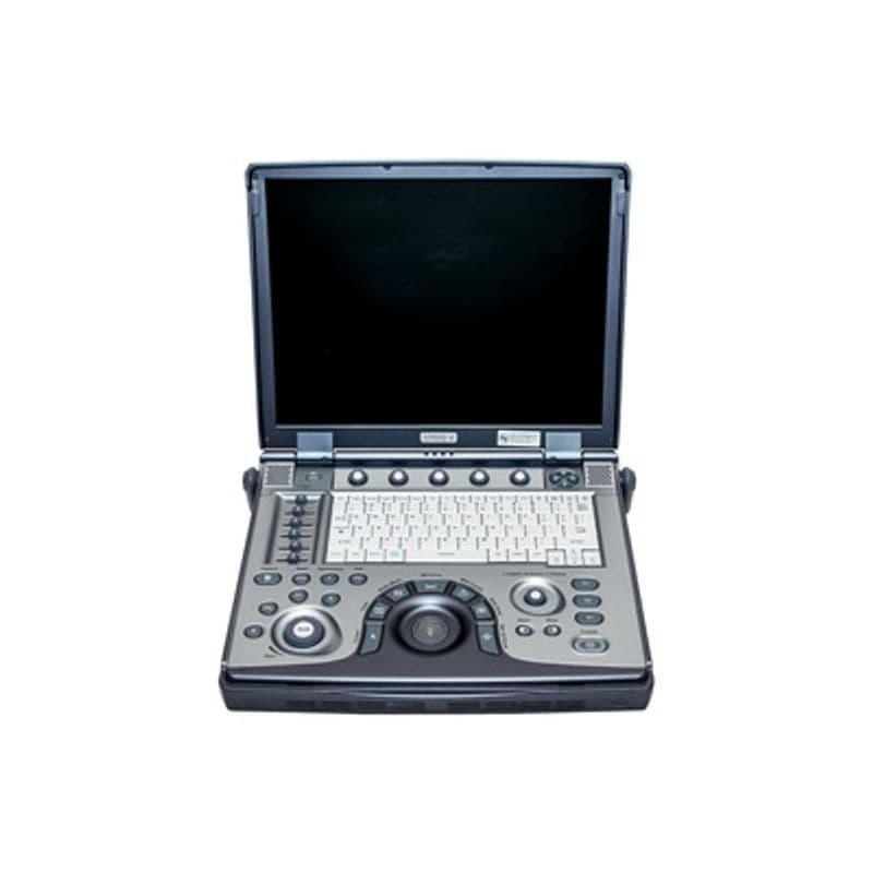
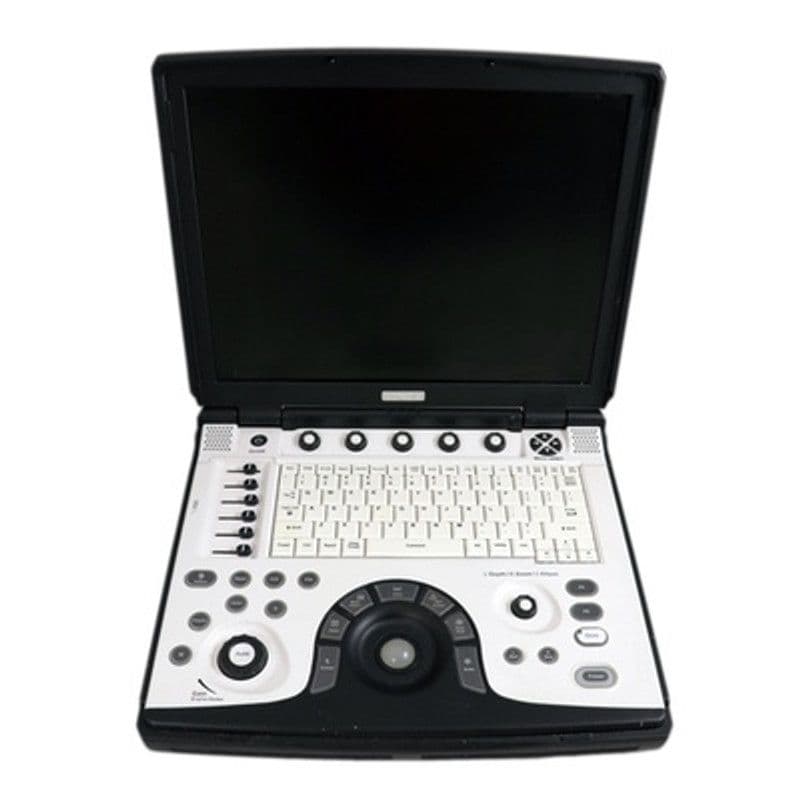
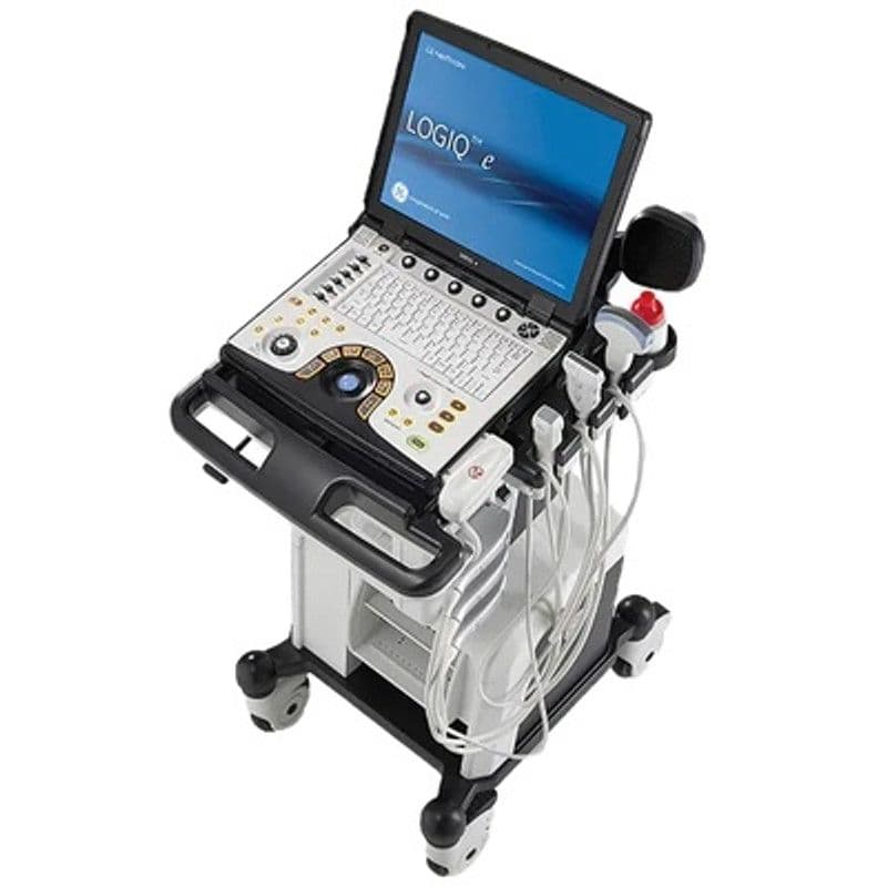
Preowned Systems in Stock
Certified preowned systems available now. Inventory changes frequently — contact us for the latest availability and pricing.
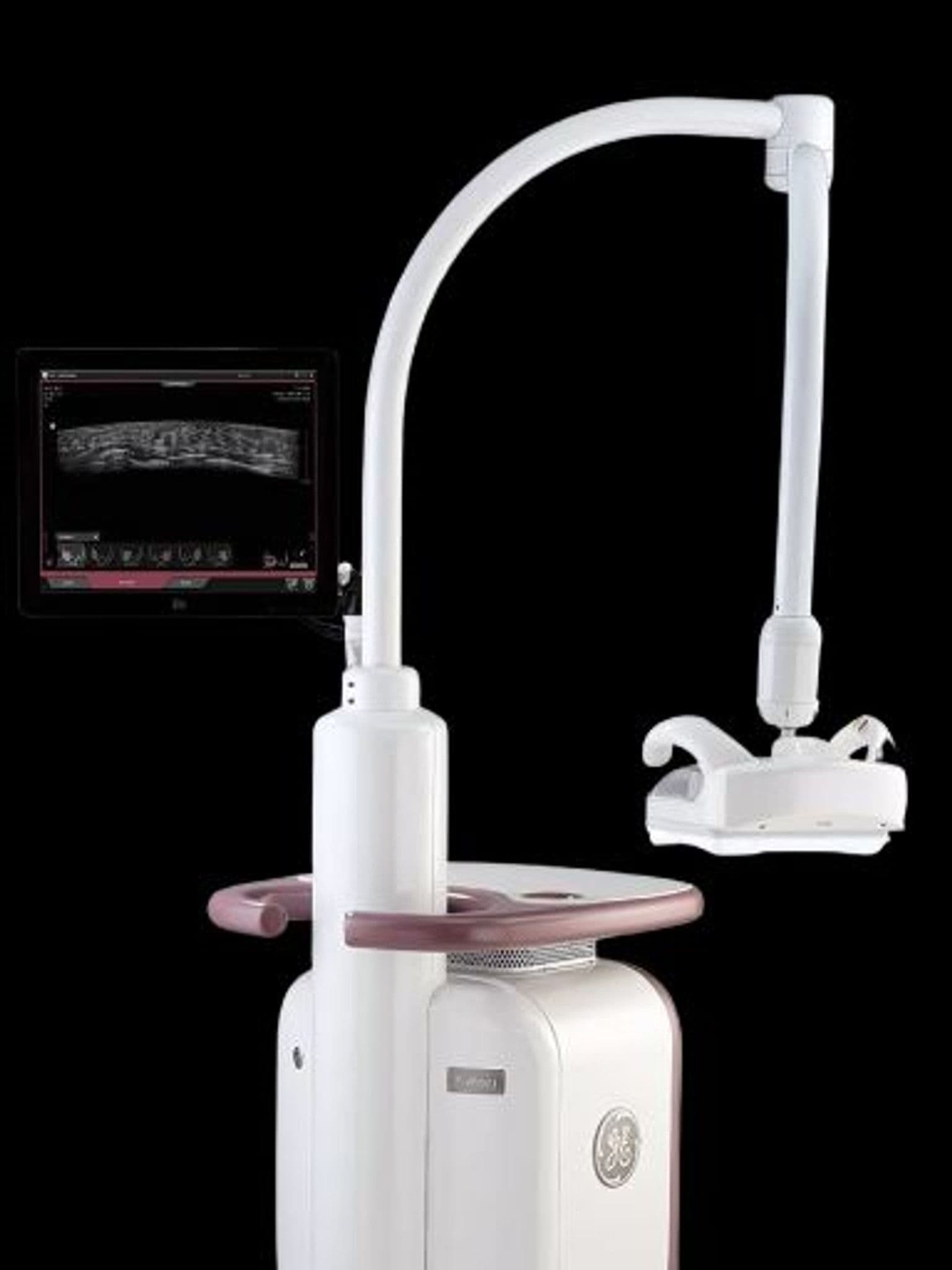
GE Invenia ABUS 2.0
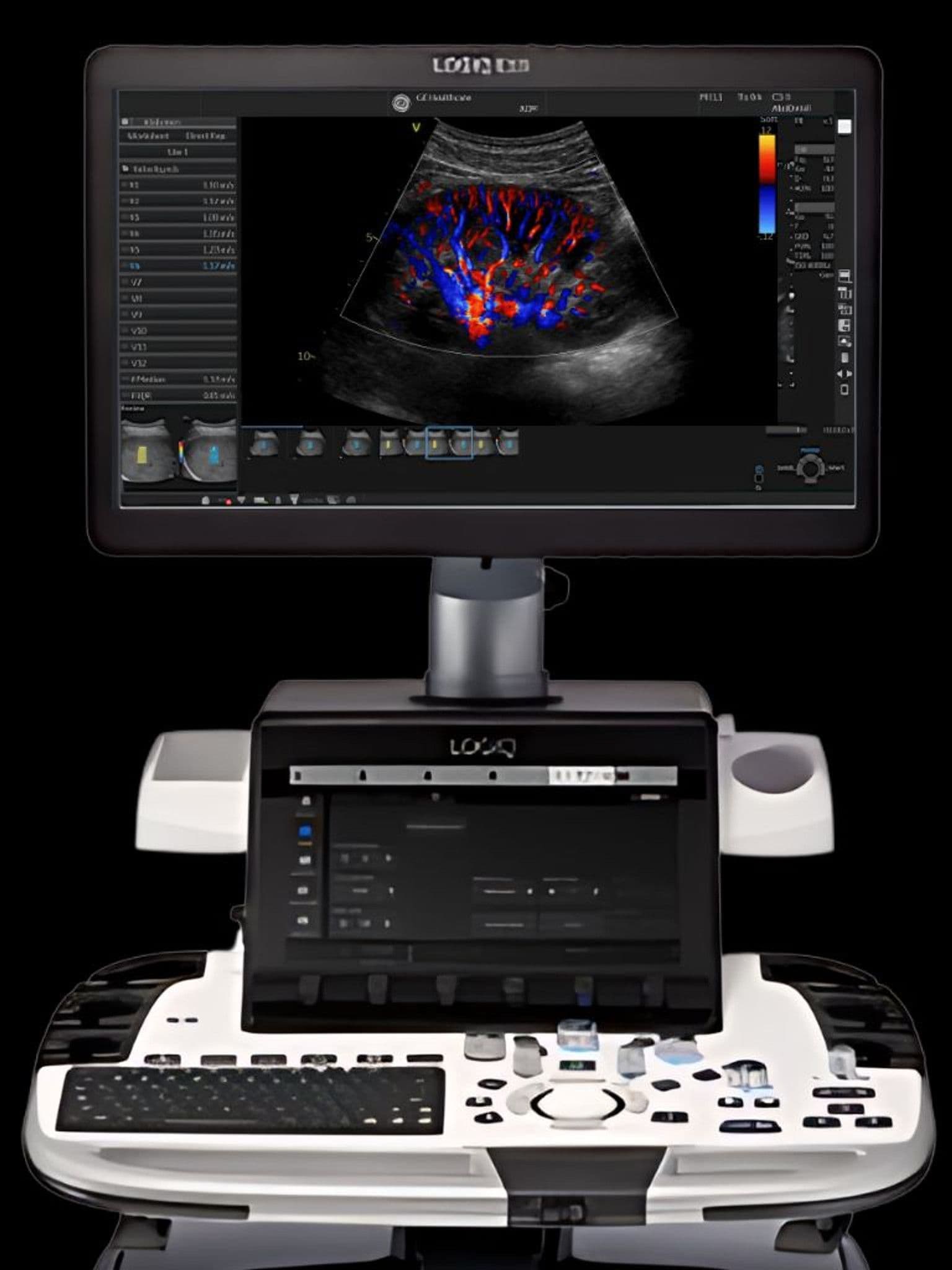
GE LOGIQ E10
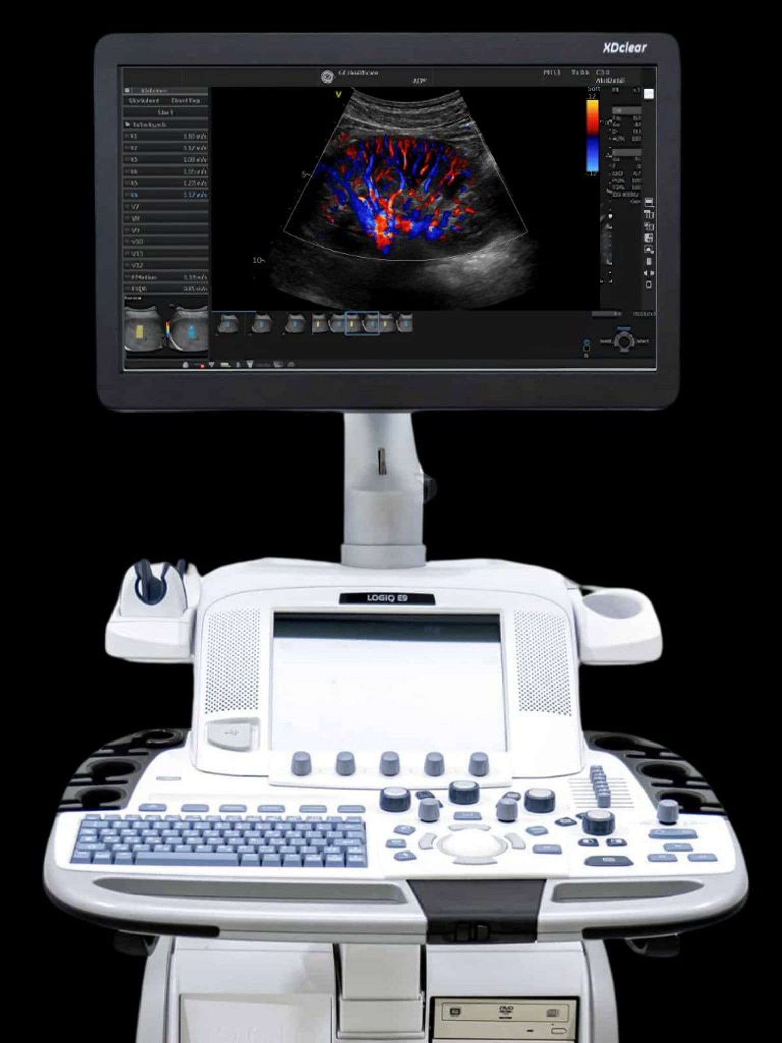
GE LOGIQ E9
GE LOGIQ F8
GE LOGIQ Fortis
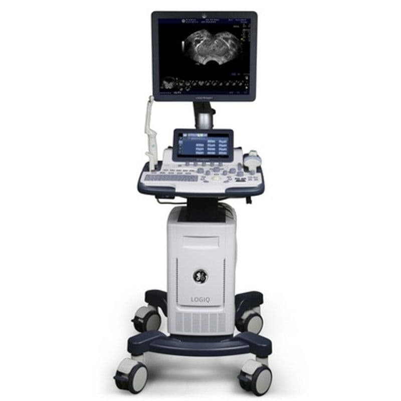
GE LOGIQ P10
GE LOGIQ P5
GE LOGIQ P9
GE LOGIQ Totus
GE LOGIQ V5
GE LOGIQ e BT11
GE LOGIQ e BT12
GE NextGen LOGIQ e R7
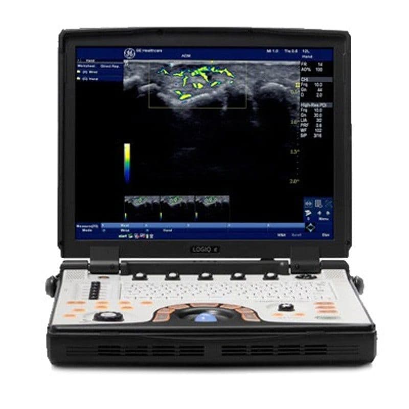
GE NextGen LOGIQ e R8
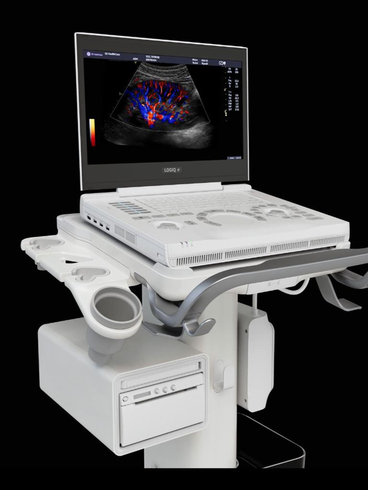
GE NextGen LOGIQ e R9
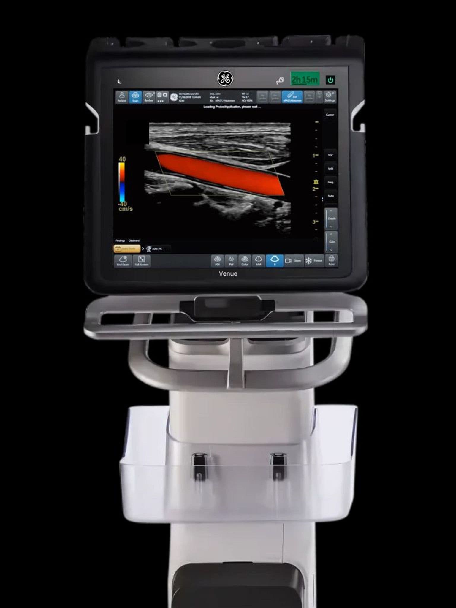
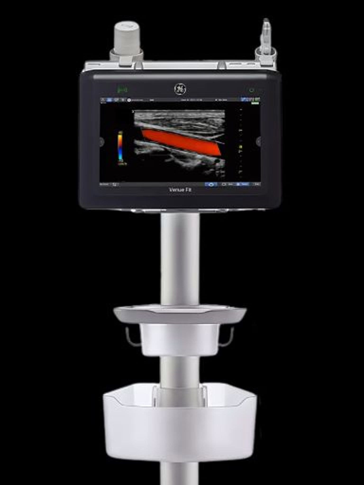
GE Venue Fit
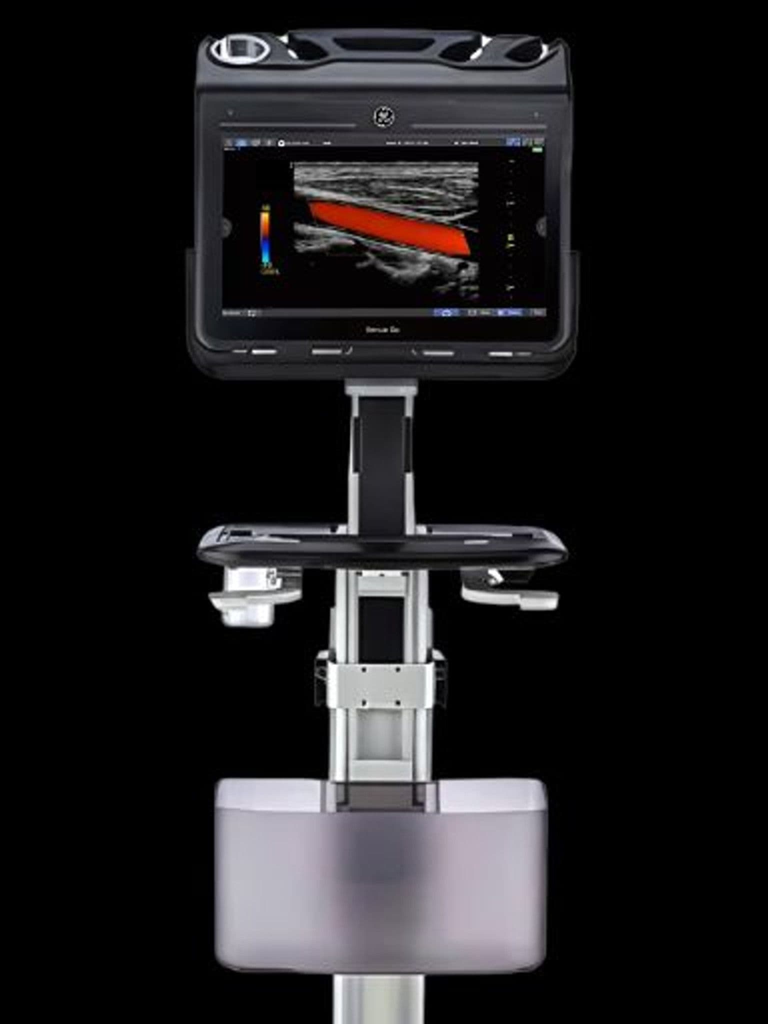
GE Venue Go
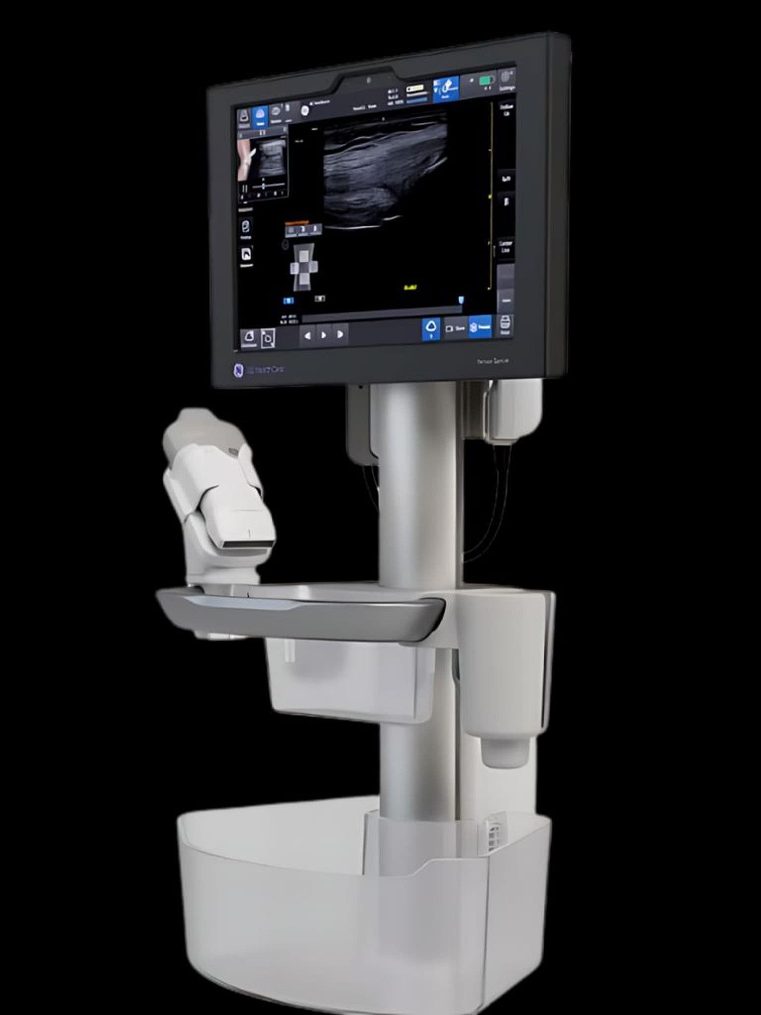
GE Venue Sprint
GE Versana Active
GE Versana Balance

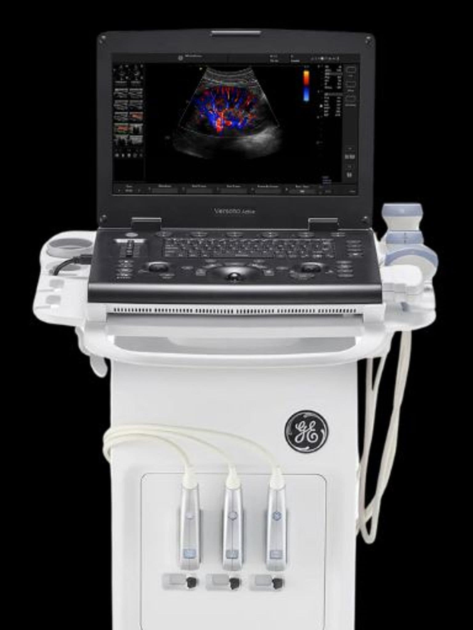
GE Versana Essential
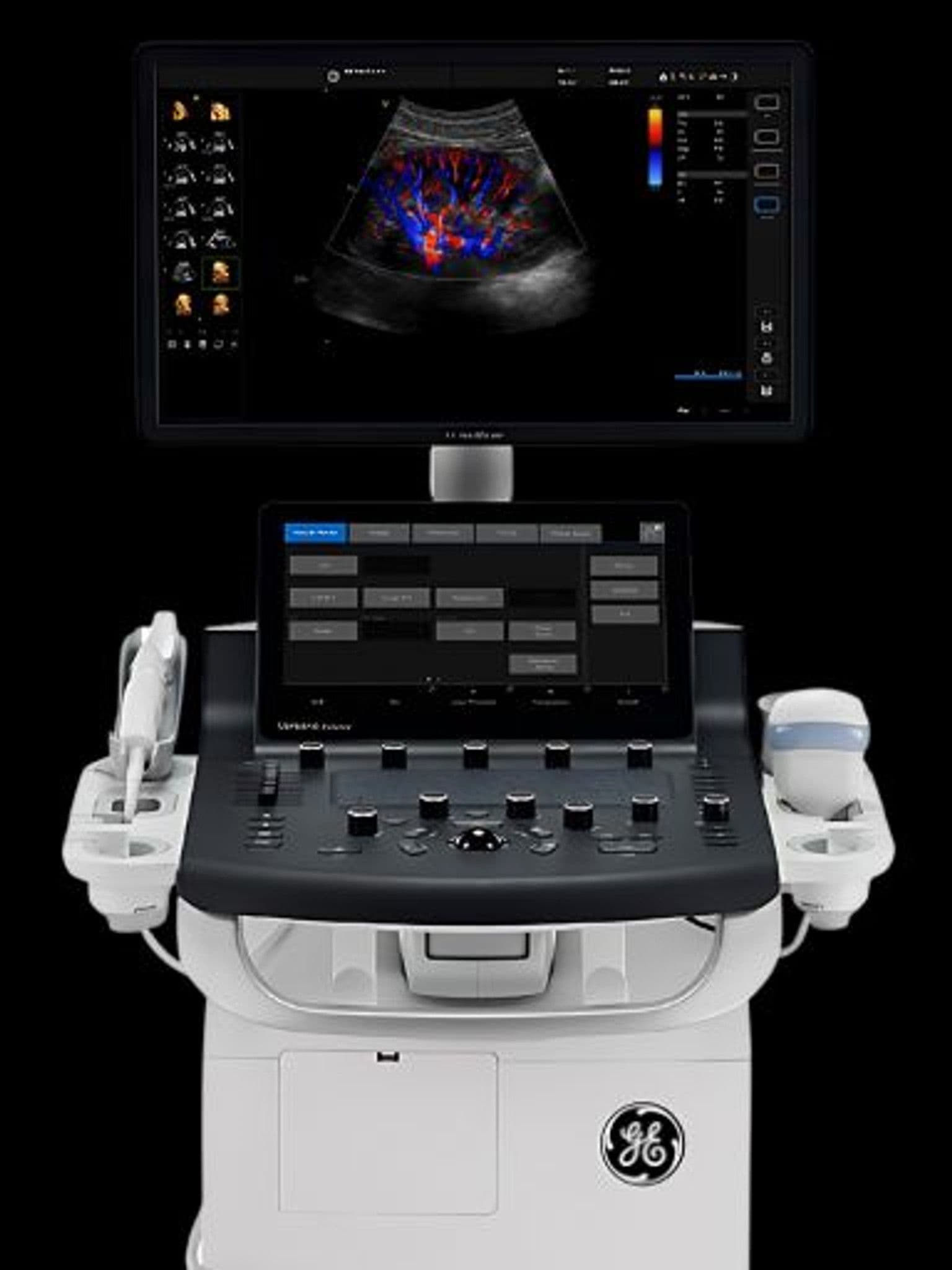
GE Versana Premier
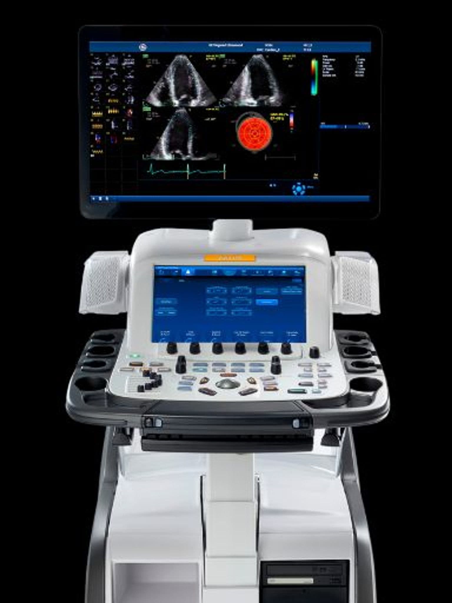
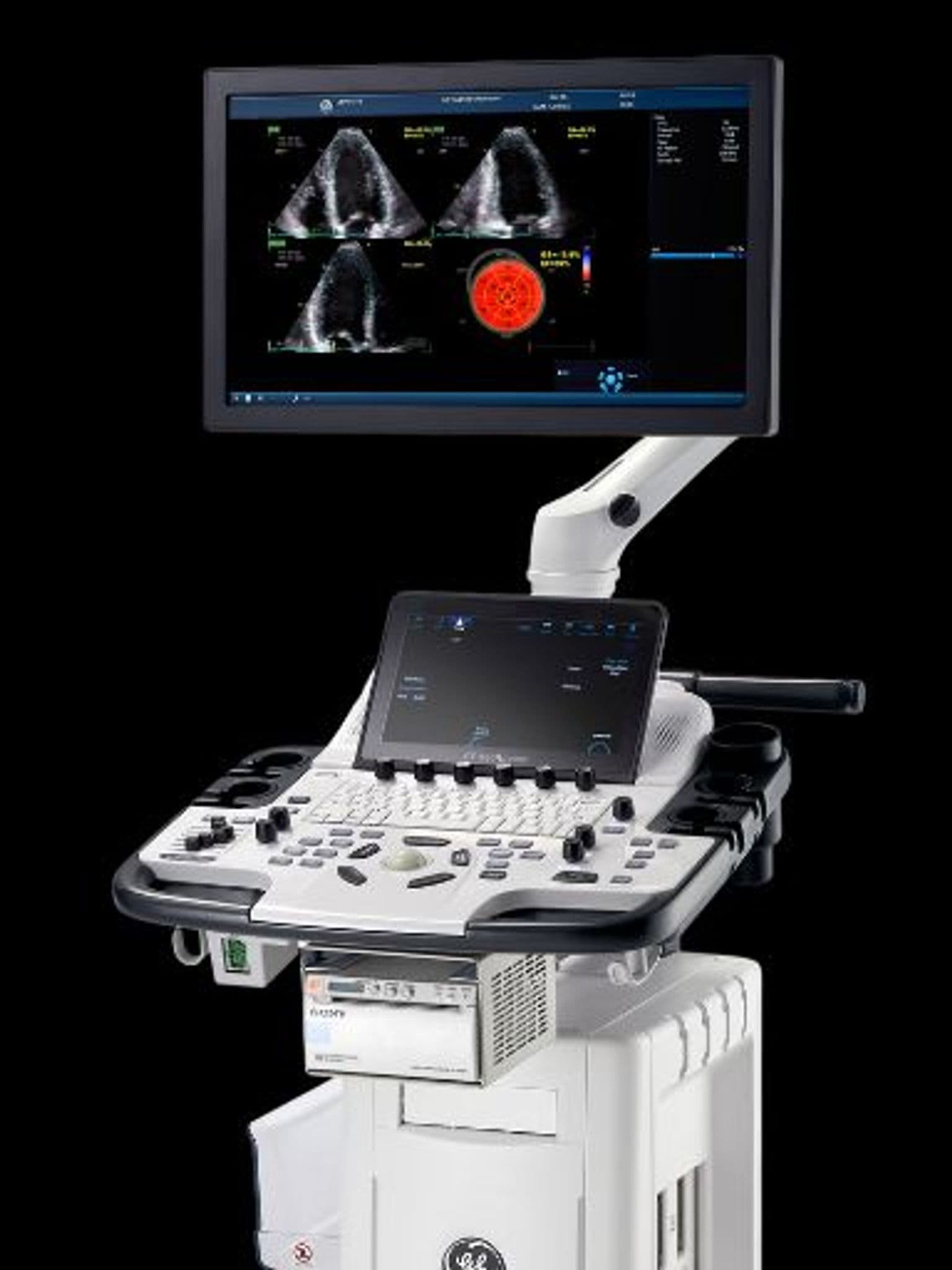
GE Vivid E95
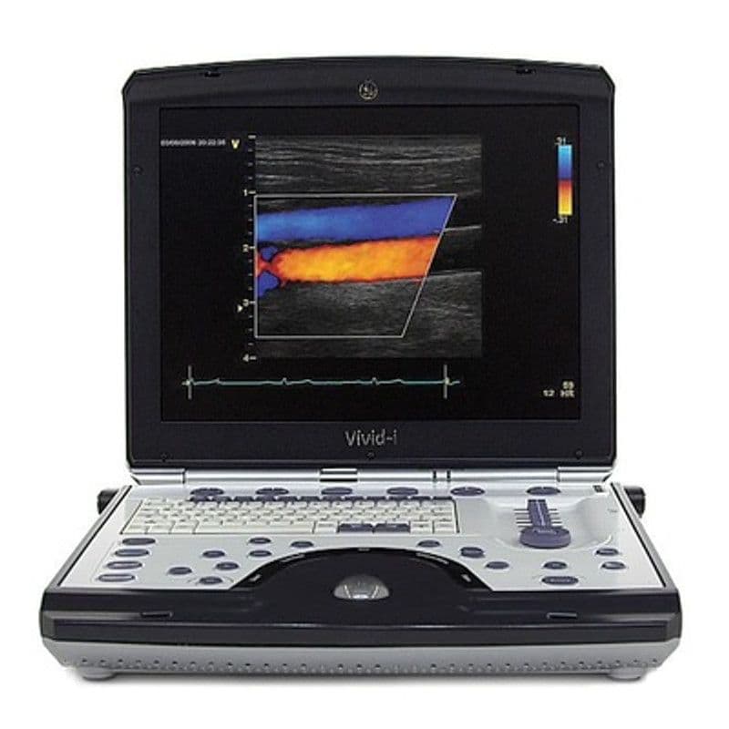
GE Vivid I BT12
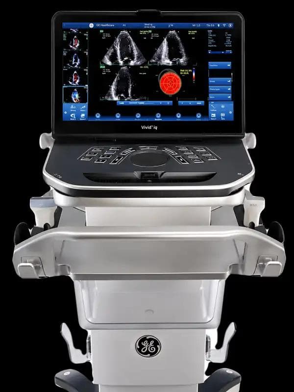
GE Vivid IQ

GE Vivid S70N
GE Vivid T9
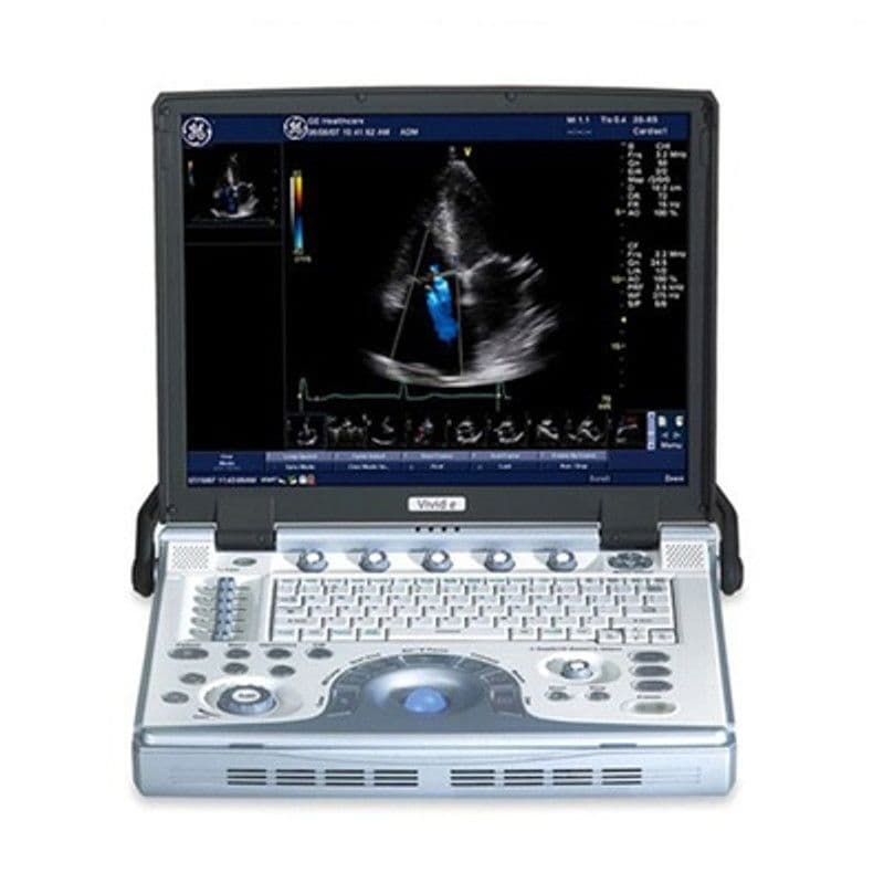
GE Vivid e
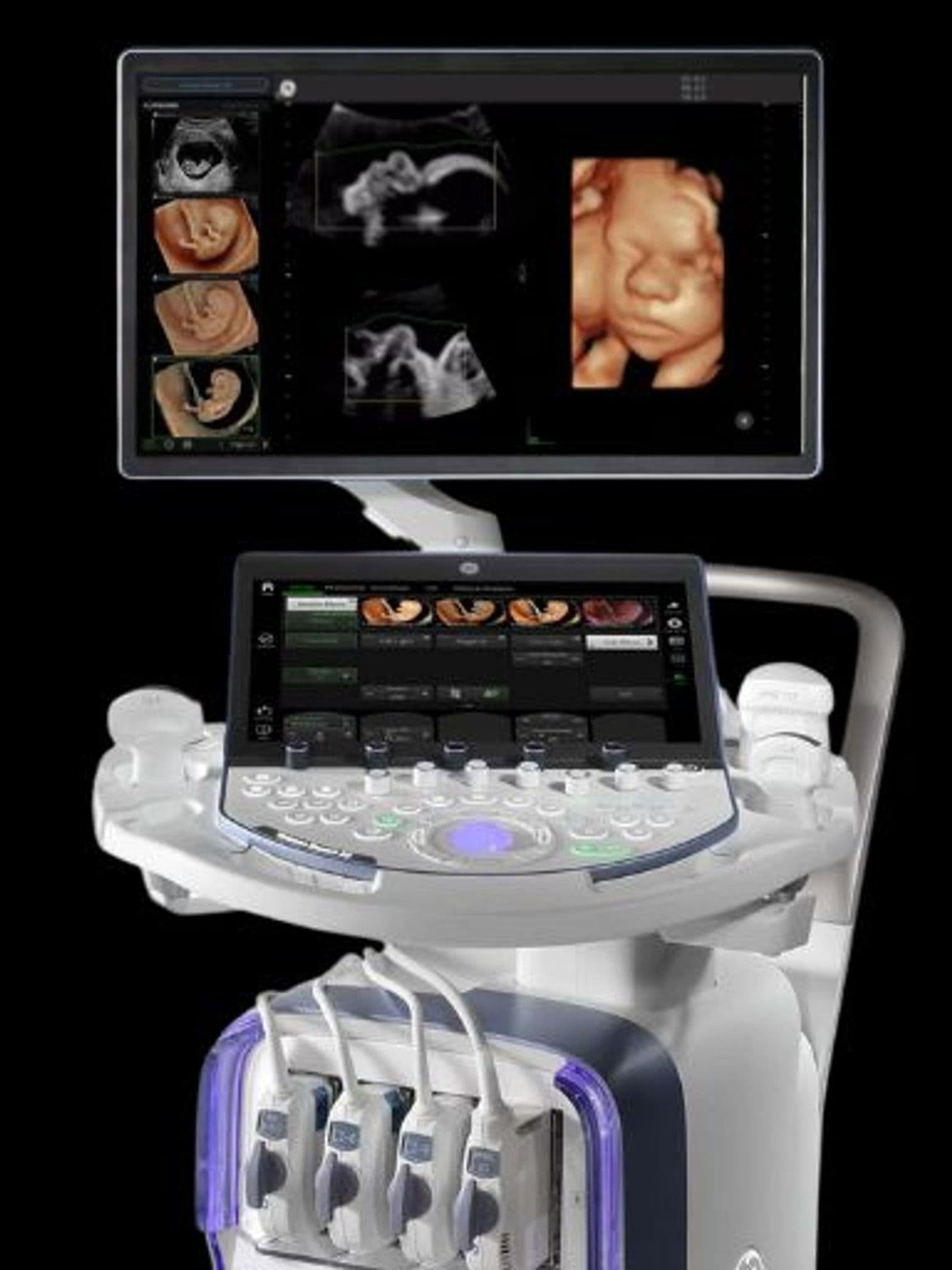
GE Voluson Expert 22

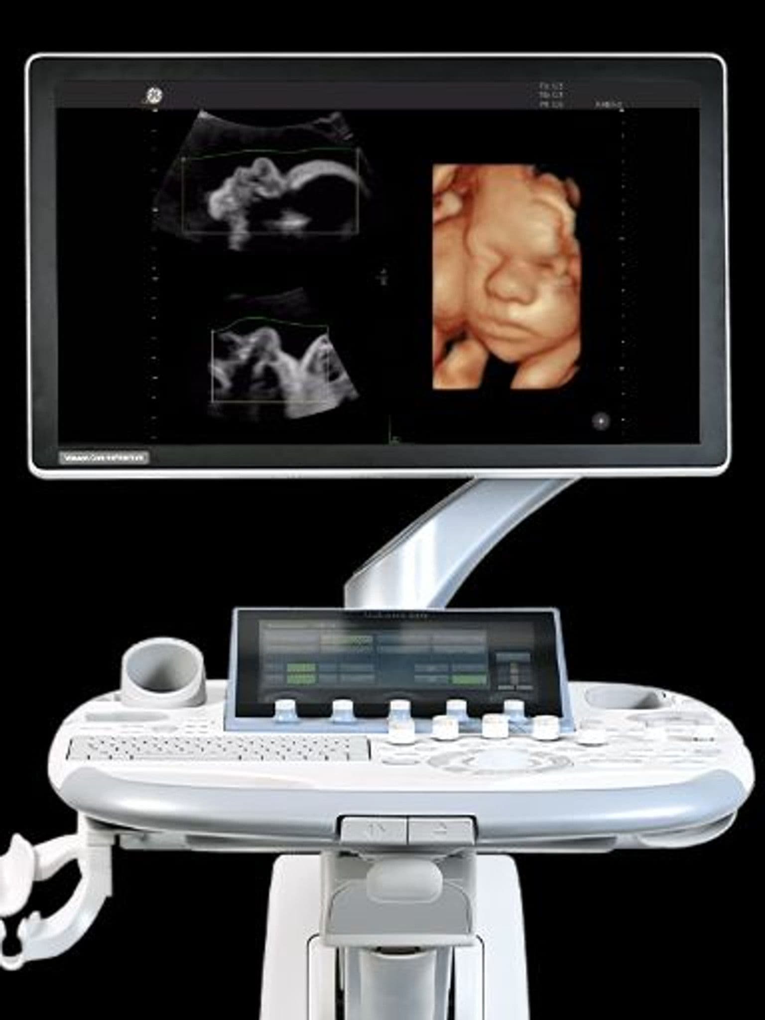
GE Voluson P8
GE Voluson S10 Expert
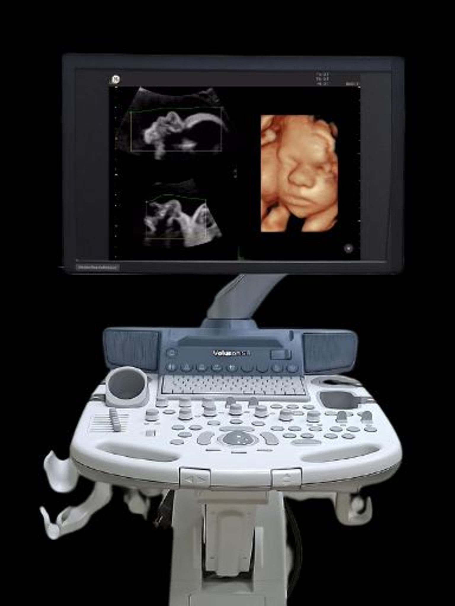
GE Voluson S8
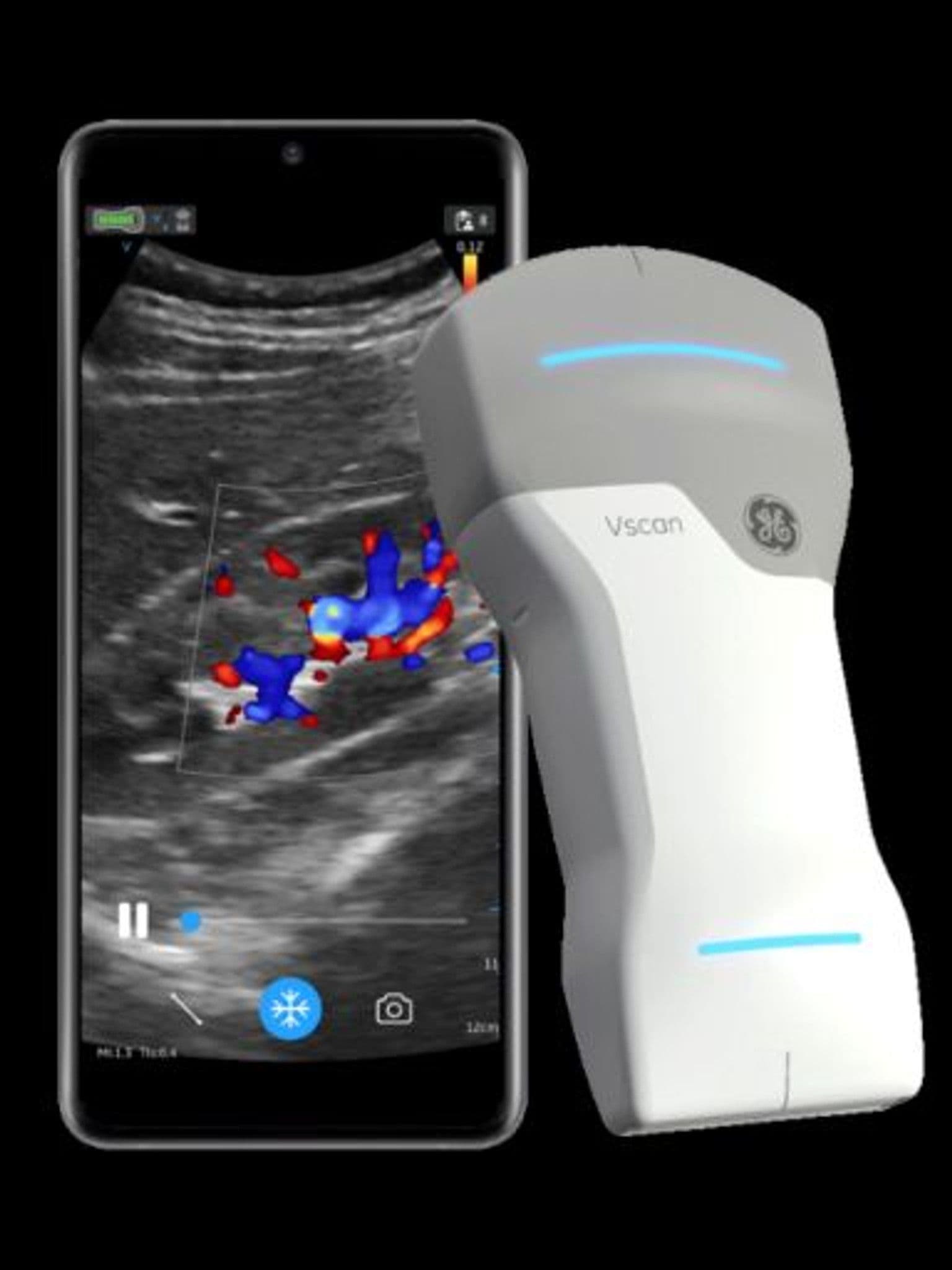
GE Vscan Air
Our Partners


Get preowned pricing for your specialty
Tell us your specialty and patient volume. We'll recommend the best preowned options with pricing and warranty details.